Authors: Tae An Choi ANP-BC & Back Kim MD FACC.
Heart Vein NYC, New York, New York

A 56-year-old female, suffering chronic leg pain, heaviness, fatigue, achiness and swelling, with associated pelvic/gluteal/hip discomfort, worsening since provoked DVT of left lower extremity, status post hysterectomy 14 years ago.
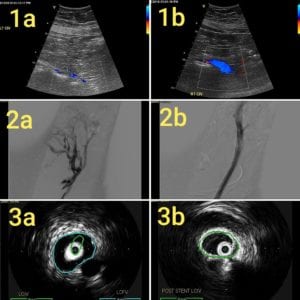
Pre-stent sonogram Doppler (1a) showing narrowing of left common iliac vein compression (CIV). Post-stent sonogram Doppler (1b) showing unobstructed venous flow of left CIV.
Pre-stent venogram (2a) showing near-occlusion of left common and external iliac vein with collateral veins. Post-stent venogram (2b) demonstrating unobstructed venous outflow and resolution of collateral flow.
Pre-stent intravascular ultrasound (IVUS) (3a) demonstrating compression of the left common iliac vein (CIV) by the right common iliac artery (CIA). Post-stent IVUS (3b) showing optimal decompression of left CIV.
At 1 week Post-stent follow-up, ultrasound showed patent stents without evidence of thrombosis. Almost complete resolution of leg and gluteal symptoms with significantly reduced left leg swelling.


